Apr 10, 2024
Ventral slot surgery is a commonly used surgical approach in dogs with cervical disc disease to decompress the spinal cord, alleviate pain, and prevent worsening of neurological signs. This procedure can be complicated and is best performed by a well-trained and experienced neurosurgery team.
Intervertebral disc disease (IVDD) is a neurologic condition that involves the vertebrae, intervertebral discs, spinal cord, and nerves branching off the spinal cord. Intervertebral discs are composed of a thick fibrous outer layer and a jelly-like inner center, and they normally act as a cushion between the vertebrae. IVDD causes the intervertebral disc to bulge or burst into the space around the spinal cord, which can cause pain, nerve damage, and paralysis.
Cervical disc disease (i.e., a slipped disc in the neck) occurs when IVDD affects the intervertebral discs in the neck region. Signs may include:
Cervical disc disease can affect any breed, but chondrodystrophic dogs (i.e., those with short legs and long bodies) are at an increased risk. Breeds most commonly affected include French bulldogs, dachshunds, beagles, poodles, shih tzus, Pekingese, and Chihuahuas.
If your pet has signs that may indicate a problem in their neck, the diagnostic process typically involves the following steps:
Surgery is often necessary to address cervical disc disease in a pet who has repeated neck pain episodes, severe neck pain, or neurologic symptoms, or who isn’t responding to conservative treatment.
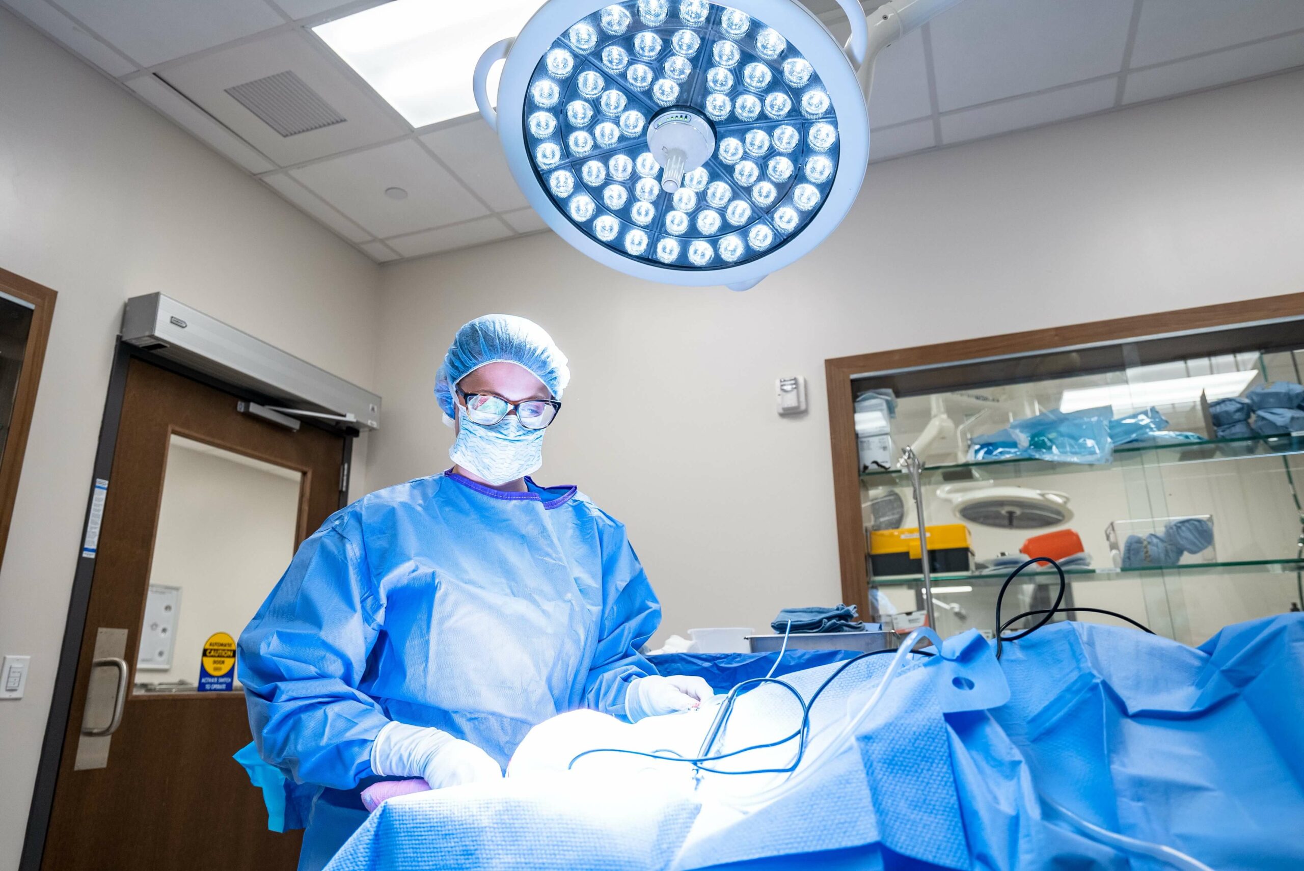
Ventral slot surgery is the most commonly performed procedure to treat disc disease in the neck. This technique involves making an incision along the midline under the neck, and retracting the soft tissues to either side so the neurosurgeon can visualize the affected disc space. Using specialized equipment, the neurosurgeon cuts a rectangular window through the affected disc and a section of the adjacent vertebrae to access the spinal canal. They gently remove the disc material compressing the spine and any remaining material inside the disc to help prevent recurrence.
After a ventral slot procedure, pets are hospitalized, typically for one to five days, with intravenous (IV) fluids and medications administered to control pain and inflammation. If the pet cannot move around on their own, the veterinary team will reposition them frequently to prevent pressure sores and other complications. Some pets may require help with urination and defecation. Once the pet can eat and drink on their own and oral medications can manage their pain, they can go home. Most pets are significantly more comfortable 24 hours after surgery, and neurologic symptoms usually improve over several days. Maximum nerve function recovery can take two weeks to several months, depending on the severity and duration of symptoms before surgery.
Once at home, pets must be strictly confined, and a chest harness should be used to avoid placing pressure on the neck. The pet must not jump, run, play, or use the stairs for the first few weeks, and take only short, slow leash walks. Raising the pet’s food and water bowls is also recommended, so they don’t have to move their neck too much to eat and drink.
Pets who have mobility issues require more intensive at-home care, including frequent repositioning, using a sling to take them for bathroom breaks, and regular bladder expression if they can’t empty their bladder on their own. Pets should also be kept clean and dry to avoid complications from urine and feces soiling.
The pet’s prognosis following a ventral slot surgery depends on the severity and duration of symptoms before surgery:
If your pet is experiencing neck pain or difficulty walking and potentially needs treatment for cervical disc disease, contact our Animal Emergency & Specialty Center neurosurgery department for expert care.